


 Media kit
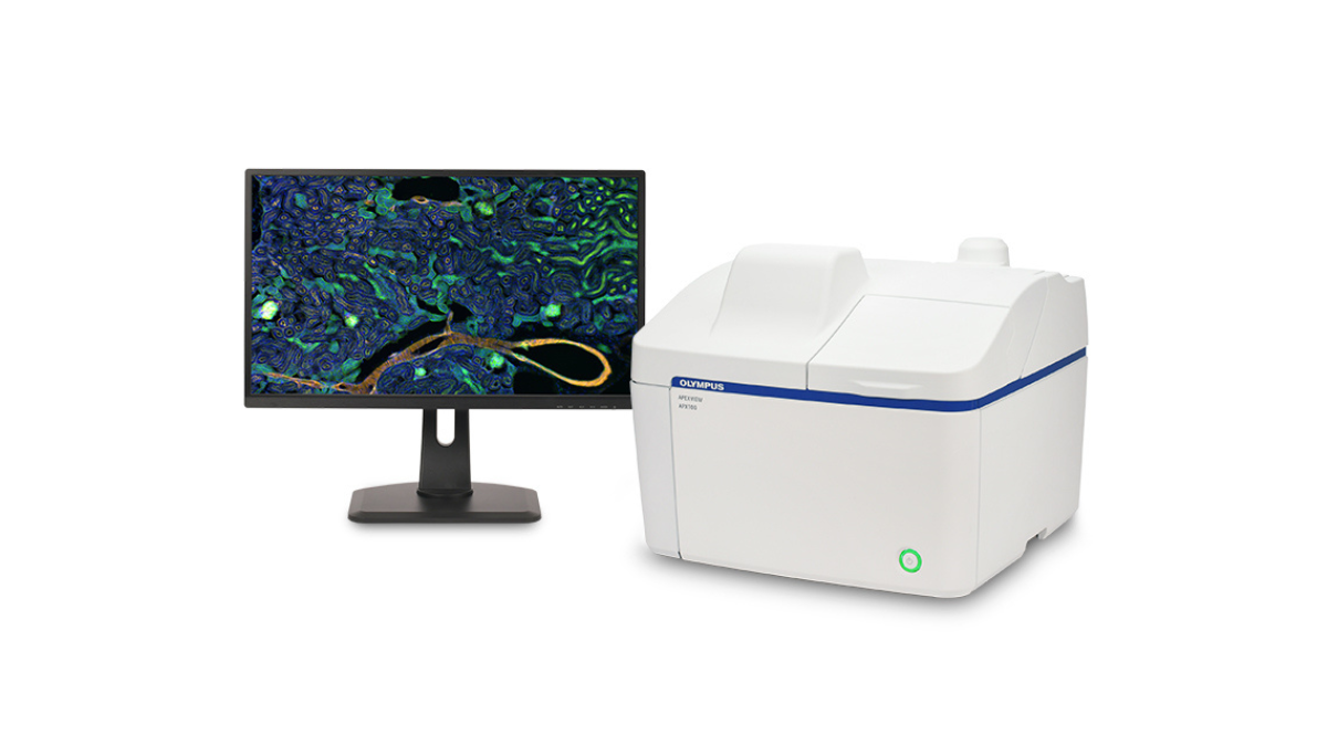
Featuring Olympus' renowned optical components, intuitive software, powerful artificial intelligence and a variety of practical functions, the APX100 system combines the ease of use of an all-in-one microscope with publication-quality images to make life science research more efficient. Designed for laboratories and imaging departments, this space-saving microscope features an integrated anti-vibration mechanism and light-shielded optical components that allow the system to be placed anywhere, even in a brightly lit room.
Simplified image production for research
While conventional microscopes may require time-consuming adjustments to capture good images, the APX100 imaging system does most of these adjustments automatically so you can focus on your research.
Using the APX100 imaging system is simple: just insert the sample, close the lid and press a button. The system then automatically acquires a preview macro image, and the built-in AI locates and displays samples on the monitor, so you can start capturing images straight away. This process is completed rapidly thanks to the system's enhanced autofocusing. It's up to twelve times faster than conventional autofocus methods, so you can quickly find the ideal imaging plane.
Produce publication-quality images in just a few clicks
The APX100 imaging system uses the same optical components as Olympus' state-of-the-art research microscopes, enabling the acquisition of publication-quality images for a wide range of applications. Most objectives are compatible with the system, including the award-winning X Line and silicone immersion objectives. They all enable high-quality image acquisition for advanced research applications.
The system also features a new gradient contrast method, which allows for sharp, high-contrast imaging of live cells or thin, unstained tissue sections without the use of specialized differential interference contrast or phase contrast optical components. This method has many unique advantages: it is less affected by meniscus, vessel lids, and water droplets, and it can be used on both glass and plastic bottomed dishes and multi-well plates. It also allows you to image through the plastic lids of Petri dishes and multi-well plates, reducing the risk of contamination.
Fast and efficient data management
To keep images in order, the APX100 system automatically organizes and stores your data. When an image is acquired, the software creates folders for each sample and saves the data in the correct folder. All important acquisition parameters are saved with the images so that they can be easily recalled for future experiments.
See the APX system in its first public presentations
- June 15-17 - International Society for Stem Cell Research (Booth #629), Moscone West Convention Center, San Francisco, California, USA
- June 21-24 - Analytica (room A2, booth #311), Messe München, Munich, Germany
- June 30 - July 2 - Japan Neuroscience Society, Okinawa Convention Center, Okinawa, Japan
- August 13-17 - Chinese Society for Cell Biology, Xiamen International Conference Center, Fujian, China


